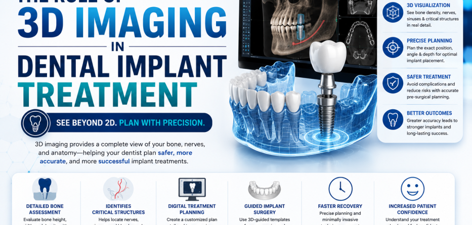
Dental implant technology has advanced significantly over the past few decades, making treatments safer, more accurate, and more predictable than ever before. One of the most important innovations driving this progress is 3D imaging technology.
Traditional dental X-rays provide valuable information, but they only show a two-dimensional view of oral structures. In contrast, 3D imaging allows dentists to visualize the teeth, jawbone, nerves, and surrounding tissues from multiple angles, creating a detailed digital model of the patient’s mouth.
This advanced imaging technology has transformed the way dental implants are planned and placed, helping improve precision, reduce complications, and enhance treatment outcomes.
In this article, we’ll explore the role of 3D imaging in dental implant treatment, its benefits, and why it has become a vital part of modern implant dentistry.
What Is 3D Imaging in Dentistry?
3D imaging refers to advanced scanning technology that captures highly detailed three-dimensional images of the teeth, gums, jawbone, and facial structures.
The most commonly used technology for dental implants is cone beam computed tomography (CBCT).
Unlike traditional X-rays, CBCT scans provide the following:
- Three-dimensional views
- Precise measurements
- Detailed bone analysis
- Nerve and sinus location mapping
- Accurate implant planning data
This information helps dentists make more informed treatment decisions.
Why Is 3D Imaging Important for Dental Implants?
Dental implant placement requires exceptional precision.
A successful implant must be positioned correctly within the jawbone while avoiding sensitive anatomical structures such as nerves, blood vessels, and sinus cavities.
3D imaging helps dentists:
- Assess bone quality
- Evaluate bone volume
- Identify anatomical landmarks
- Plan implant placement accurately
- Reduce surgical risks
The result is a safer and more predictable treatment process.
How 3D Imaging Improves Implant Planning
Comprehensive Bone Assessment
One of the most important requirements for dental implants is sufficient bone support.
3D scans allow dentists to evaluate the following:
- Bone density
- Bone width
- Bone height
- Bone defects
This helps determine whether a patient is a suitable candidate for implants or may require procedures such as bone grafting.
Accurate Implant Positioning
Proper implant positioning affects:
- Function
- Stability
- Appearance
- Longevity
Using 3D imaging, dentists can identify the ideal angle, depth, and location for implant placement.
Benefits include:
- Improved aesthetics
- Better chewing function
- Reduced complications
- Enhanced long-term success
Identification of Critical Structures
Certain areas of the mouth contain sensitive structures that must be protected during implant surgery.
These include:
- Inferior alveolar nerve
- Mental nerve
- Maxillary sinuses
- Adjacent tooth roots
3D imaging allows dentists to visualize these structures clearly and avoid accidental injury.
The Role of CBCT Scans in Implant Dentistry
What Is a CBCT Scan?
Cone Beam Computed Tomography (CBCT) is a specialized imaging system designed specifically for dental and maxillofacial applications.
The scanner rotates around the patient’s head, capturing hundreds of images that are reconstructed into a detailed 3D model.
Advantages of CBCT
- High-resolution imaging
- Quick scanning process
- Lower radiation exposure than traditional medical CT scans
- Precise treatment planning
- Enhanced diagnostic capabilities
CBCT has become the gold standard for implant assessment and planning.
Guided Implant Surgery and 3D Imaging
One of the most significant advancements in implant dentistry is guided implant surgery.
Using 3D imaging data, dentists can create the following:
- Digital treatment plans
- Virtual implant simulations
- Surgical guides
These custom guides help position implants exactly as planned.
Benefits of Guided Surgery
- Increased accuracy
- Reduced surgical time
- Less invasive procedures
- Faster healing
- Improved patient comfort
This technology significantly improves treatment predictability.
Benefits of 3D Imaging for Patients
Enhanced Safety
Detailed imaging helps identify potential risks before surgery begins.
This reduces the likelihood of the following:
- Nerve injury
- Sinus complications
- Implant misplacement
Improved Treatment Outcomes
Better planning leads to:
- Higher success rates
- Improved aesthetics
- Better implant stability
- Long-term durability
Faster Treatment Process
Accurate diagnosis and planning help streamline treatment and reduce unexpected delays.
Better Patient Education
3D images help patients visualize their treatment plan and understand the procedure more clearly.
How 3D Imaging Helps with Complex Cases
Certain implant cases require advanced planning.
Examples include:
Full-Arch Implant Restorations
Patients replacing multiple teeth need precise implant placement to support full-arch prosthetics.
Bone Loss Cases
3D imaging helps evaluate the extent of bone loss and determine whether bone grafting is necessary.
Sinus Lift Procedures
For implants in the upper jaw, CBCT scans help assess sinus anatomy and guide treatment planning.
Immediate Implant Placement
When implants are placed immediately after extraction, precise imaging is critical for success.
Comparing Traditional X-Rays and 3D Imaging
| Feature | Traditional X-Rays | 3D Imaging (CBCT) |
|---|---|---|
| View | 2D | 3D |
| Bone Analysis | Limited | Detailed |
| Implant Planning | Basic | Highly Accurate |
| Nerve Visualization | Limited | Excellent |
| Surgical Precision | Moderate | High |
| Treatment Predictability | Moderate | Excellent |
This comparison demonstrates why 3D imaging has become a standard tool in implant dentistry.
Does 3D Imaging Increase Implant Success Rates?
While many factors contribute to implant success, 3D imaging significantly improves treatment planning and surgical accuracy.
Benefits that contribute to higher success rates include:
- Better implant positioning
- Reduced surgical errors
- Improved bone assessment
- Enhanced patient selection
These advantages help create more predictable outcomes and long-term implant stability.
Future Trends in 3D Imaging and Implant Dentistry
Dental technology continues to evolve rapidly.
Emerging innovations include:
- Artificial intelligence-assisted planning
- Digital smile design integration
- Fully guided implant surgery
- Real-time surgical navigation
- Enhanced digital workflows
These technologies are expected to further improve implant precision and patient outcomes.
Frequently Asked Questions
Is 3D imaging necessary for dental implants?
While not mandatory in every case, 3D imaging provides critical information that improves planning, accuracy, and safety.
What is the difference between a CBCT scan and a regular dental X-ray?
CBCT scans provide a three-dimensional view of oral structures, while traditional X-rays offer only a two-dimensional image.
Is a CBCT scan safe?
Yes. CBCT scans use relatively low radiation doses and are considered safe when used appropriately.
How long does a 3D dental scan take?
Most CBCT scans take less than one minute to complete.
Can 3D imaging detect bone loss?
Yes. CBCT technology provides highly accurate measurements of bone volume and density.
Conclusion
The role of 3D imaging in dental implant treatment cannot be overstated. By providing detailed, three-dimensional views of the jawbone, teeth, nerves, and surrounding structures, this technology allows dentists to plan and place implants with remarkable precision.
From improving safety and accuracy to enhancing patient outcomes and treatment predictability, 3D imaging has become an essential tool in modern implant dentistry. As technology continues to evolve, advanced imaging systems will play an even greater role in delivering successful, long-lasting dental implant results.